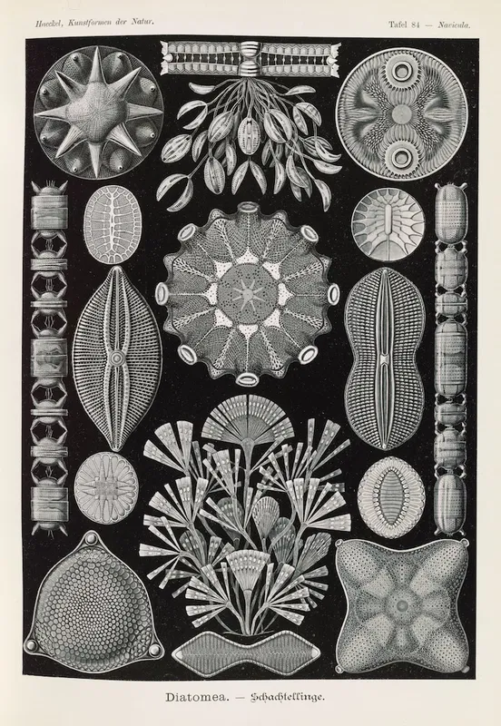
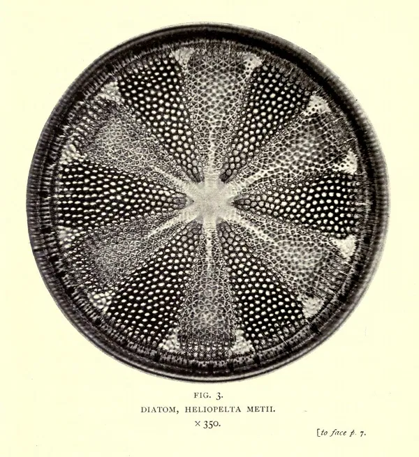
Microscopic yet mighty, diatoms are photosynthetic microorganisms that are all around us. These hardy organisms are found in nearly every aquatic environment on Earth, including freshwater, brackish, and marine ecosystems. Diatoms exhibit staggering diversity, with an estimated 100,000-200,000 distinct species. While they are well known for their incredible morphological and ecological range, they all share one very important characteristic: their porous silica frustule.
Diatoms could be a crucial next step in cancer research, drug development and delivery. A major challenge in cancer treatment is targeting cancer-affected cells with an apoptosis-causing drug while sparing surrounding noncancerous cells. Radiation therapy, for example, essentially blasts the affected area and nearby tissue in order to prevent metastasis. There has been some success with synthetic nanoparticle drug delivery systems; however, these nanoparticles are costly and time-consuming to produce, making large-scale, viable treatments difficult.
.jpg)
Utilizing diatoms, as it turns out, could provide an eco-friendly and cost-effective source of nano-porous silica. They are composed of a highly organized, porous silica shell called a frustule. Researchers can isolate this outer silica layer and use it as a silica-based nanoparticle drug carrier, coating or loading it with a cancer treatment drug. The surface can then be chemically engineered so that overexpressed receptors on cancer cells recognize attached ligands on the nanoparticle and bind to it. While the silica particles themselves are generally biocompatible, when concentrated in cancer cells, they can deliver apoptosis-inducing drugs directly where they are needed. This would make cancer treatments more effective and ideally result in fewer side effects for patients.
These abundant organisms, often overlooked, could be a critical turning point in drug therapies. However, there are still challenges. Research in this area is relatively new, and the stability of the silica nanoparticles needs to be improved, as well as better control over the size and morphology of diatoms when grown in a lab. Another obstacle is the question of biodegradability, ensuring that the frustules do not accumulate in the liver, kidneys, and other vital organs and cause additional complications. Additionally, more research must be conducted to ensure drug loading efficiency and reliable drug release before this treatment tool can be clinically implemented on a larger scale.